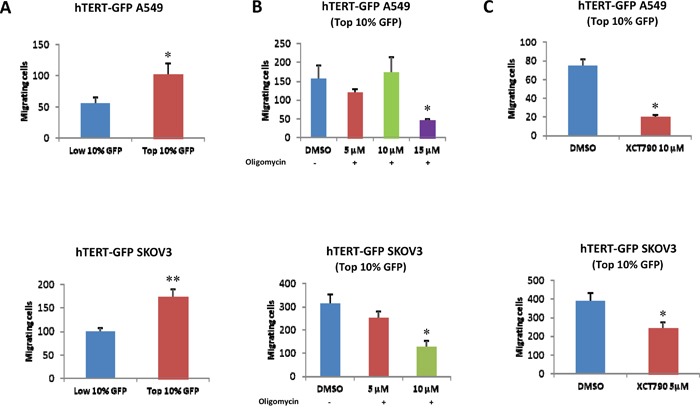
Figure 8. hTERT-high cells show an increased capacity for cell migration, which is strictly dependent on mitochondrial function.

A. Cell migration. A549 and SKOV3 cells stably-transduced with the hTERT-eGFP reporter were subjected to flow cytometry to isolate the GFP(+) and GFP(-) cell populations. Then, the migratory capacity of these two cell populations was assessed using a modified “Boyden Chamber” assay. More specifically, the cells were allowed to migrate across an 8 μm pore uncoated membrane for 12-16 hours. Note that the GFP(+) cell population (derived from A549 or SKOV3 cells) shows a near 2-fold increase in migration, as compared with the GFP(-) cells. p<0.05 for the A549 and p<0.001 for the SKOV3 cells (Student's t-test). B. Effects of mitochondrial inhibitors. To assess whether mitochondrial function is involved in cancer cell migration, mitochondrial inhibitors (either oligomycin A or XCT790) were placed in both the upper and lower chambers. Note that oligomycin A and XCT790 both effectively inhibited migration in the GFP(+) cell population. Results are shown as the mean ± SEM, t-test, two-tailed test. *p < 0.05.
