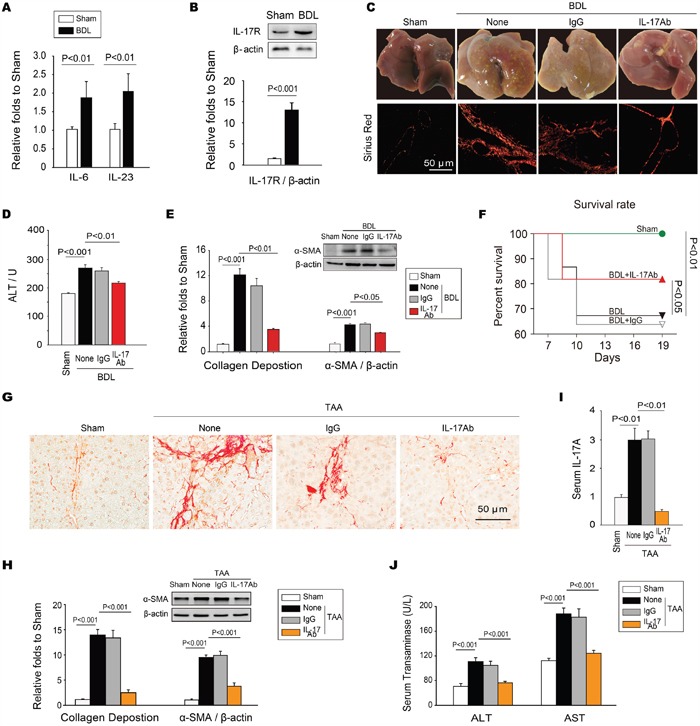
Figure 1. Neutralization of IL-17A inhibits the development of hepatic fibrosis.

A. Expressions of IL-6 and IL-23 were significantly increased in fibrotic liver tissue of the BDL-induced hepatic fibrosis. B. The IL-17R was also up-regulated in fibrotic liver tissues. C. Neutralizing IL-17A resolved BDL-induced cirrhosis. Data are morphology of the livers (top panels), as well as sirius red staining of representative photomicrographs of liver sections (bottom panels). D. Neutralization of IL-17A decreased the level of ALT in serum. E. Neutralizing IL-17A decreased the BDL-induced collagen deposition measured with sirius red staining, as well as the expression of α-SMA in hepatic tissues. F. Blocking IL-17A increased the survival rate of BDL-treated mice. The survival rate was analyzed by the Kaplan-Meier method (n=30/group). Neutralizing IL-17A also ameliorated TAA-induced chronic hepatic fibrosis. G. The representative photomicrographs of sirius red staining of the liver sections, H. the summary of collagen deposition and α-SMA expression, as well as the levels of IL-17A I., ALT and AST J. in serum indicated that neutralizing IL-17A could also ameliorate TAA-induced chronic hepatic fibrosis. Data are mean ± SEM (n=8 group-1 experiment-1) of three independent experiments.
