
Figure 3. Casein Kinase 1 (CK1) promotes FNIP2 ubiquitination and degradation.
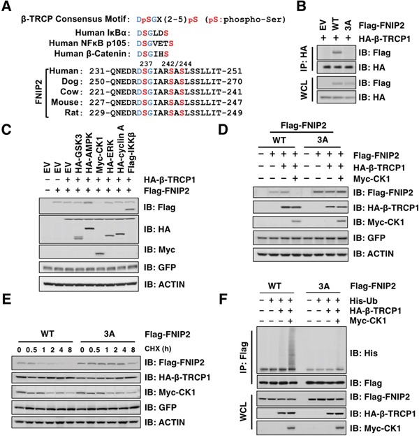
A. Alignment of FNIP2 sequences surrounding a putative β-TRCP degron motif from different species. B. Immunoblot (IB) analysis of whole cell lysates (WCL) and HA-immunoprecipitates (IP) derived from HeLa cells transfected with HA-β-TRCP1 along with empty-vector (EV) or the indicated Flag-FNIP2 constructs (WT or 3A: S237/242/244A). At 24 h post-transfection, cells were treated with MG132 for 12 h before harvesting. C. IB analysis of WCL derived from 293T cells transfected with HA-β-TRCP1 and Flag-FNIP2 along with each expression plasmids of protein kinases as indicated. GFP was included as the internal control for transfection efficiency. D. IB analysis of WCL derived from HeLa cells transfected with EV, Flag-FNIP2 (WT or 3A: S237/242/244A), HA-β-TRCP1, and Myc-CK1 as indicated. GFP was included as the internal control for transfection efficiency. E. IB analysis of WCL derived from HeLa cells transfected with Flag-FNIP2 (WT or 3A: S237/242/244A), HA-β-TRCP1, and Myc-CK1 as indicated. At 48 h post-transfection, cells were treated with 100 μg/mL cycloheximide (CHX) and harvested at the indicated time points. GFP was included as the internal control for transfection efficiency. F. IB analysis of WCL and anti-Flag IP derived from 293T cells transfected with EV, Flag-FNIP2 (WT or 3A: S237/242/244A), His-Ub, HA-β-TRCP1, and Myc-CK1 as indicated. At 24 h post-transfection, cells were treated with MG132 (15 μM) for 12 h before harvesting.