
Figure 4. β-TRCP knockdown leads to FNIP stabilization, increased FLCN lysosomal localization, and diffused mTOR distribution in HeLa cells.
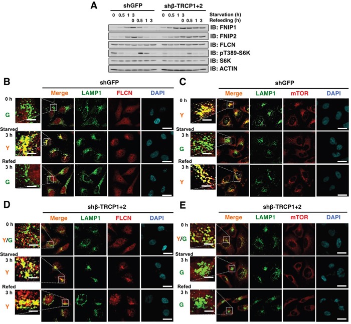
A. Immunoblot (IB) analysis of whole cell lysates derived from HeLa cells stably expressing shRNA against GFP (control) or β-TRCP1+2 (shRNA against both β-TRCP1 and β-TRCP2). After being serum and amino acid-starved for 3 h, cells were treated with fresh 10% FBS DMEM and harvested at the indicated time points. B-E. Confocal images of HeLa cells presented in (A). DAPI-loaded HeLa cells were analyzed for co-localization of FLCN (B and D) (red) or mTOR (C and E) (red) with a lysosomal marker, LAMP1 (green). Y (yellow) indicates predominant localization of FLCN or mTOR in the lysosome. Scale bars, 20 μm (5 μm in the enlarged images).