
Figure 7. Non-degradable FNIP2 mutant significantly suppresses the proliferative and tumorigenic capacity of UOK-257-2 cells.
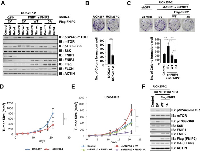
A. Immunoblot (IB) analysis of whole cell lysates derived from UOK-257-2 cells infected with shRNA lentiviral plasmids against FNIP1 and FNIP2 followed by selection in hygromycin and puromycin containing medium for 72 h, and subsequently transfected with empty vector (EV) or Flag-FNIP2 expression plasmids (WT or 3A: S237/242/244A). At 12 h post-transfection, cells were treated with 0.4 mg/mL G418 for 7 days to eliminate non-transfected cells. After being serum-starved for 3 h, cells were treated with fresh 10% FBS DMEM for 3 h before harvesting. B. Clonogenic colony formation of UOK-257 and UOK-257-2 cells (upper panel) was quantified (lower panel). Data are presented as means ± SD, n = 3, ** p < 0.01. C. Clonogenic colony formation of UOK-257-2 cells presented in Figure 7A (upper panel) was quantified (lower panel). Data are presented as means ± SD, n = 3, ** p < 0.01. D. Growth curve of developed tumors derived from xenografted UOK-257 or UOK-257-2 cells. The indicated tumor cells (3 × 10⁶) were subcutaneously inoculated into each flank of 6 nude mice. The size of visible tumors was measured at the indicated days post-injection. Data are presented as means ± SD, n = 6, ** p < 0.01. E. Growth curve of developed tumors derived from xenografted UOK-257-2 cells presented in Figure 7A or 7C. The indicated tumor cells (3 × 10⁶) were subcutaneously inoculated into each flank of 6 nude mice. The size of visible tumors was measured at the indicated days post-injection. Data are presented as mean ± SD, n = 6, ** p < 0.01. F. IB analysis of whole cell lysates derived from dissected solid tumors presented in E.