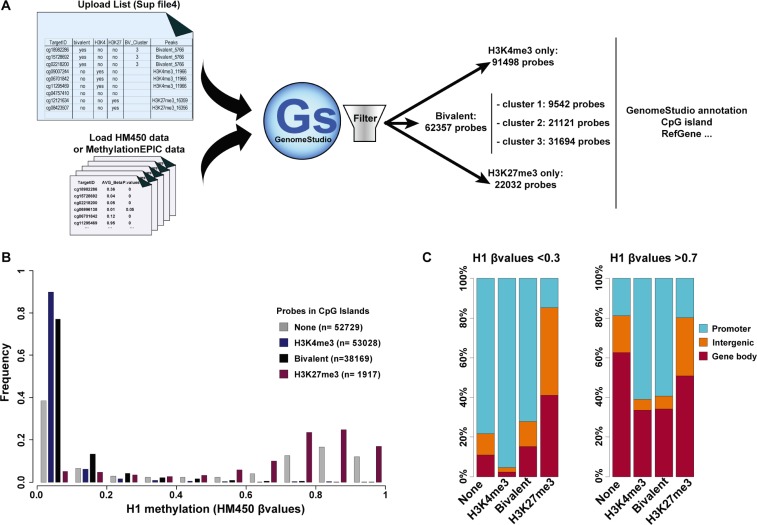
Figure 5. Integration of the ES cell chromatin signature parameters in DNA methylation analyses.
(A) Schematic overview of the approach to integrate hESC chromatin signature parameters in HM450K and MethylationEPIC (Illumina) array-based DNA methylation analyses. (B) Methylation status of CGIs in H1 hESCs according to their chromatin signature; n, number of probes per chromatin signature category. (C) Genomic features associated with unmethylated (β value < 0.3) and methylated (β value > 0.7) probes in H1 hESC CGIs according to their chromatin signature.

