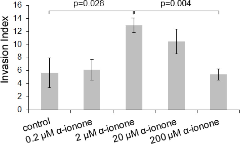
Figure 5. LNCaP cell invasiveness induced by α-ionone.
LNCaP cell invasiveness was assessed on collagen I gel in the presence of various concentrations of α-ionone (0.2, 2, 20 or 200 μM) or of 0.1% DMSO (the amount of DMSO used to dilute α-ionone before adding it to the collagen gel or culture medium). The invasion index corresponds to the percentage of cells presenting invasive extensions into the collagen gel 24 hours after seeding. Bars indicate standard deviation (n = 3). Statistics were performed using a two-tailed Student's test.

