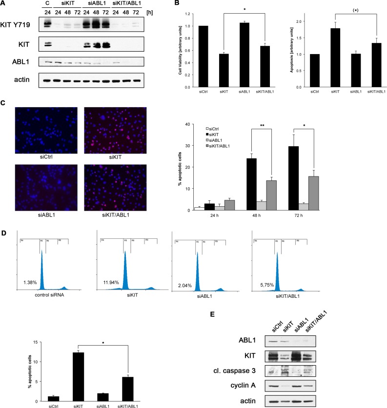
Figure 2. Co-depletion of KIT and ABL1 attenuates the effects of KIT knock-down.
(A) GIST882 cells were transfected with non-targeted siRNA control sequences (“C”) or small interfering RNA (siRNA) sequences targeting KIT and ABL1 either alone or in combination. Whole cell lysates obtained 24, 48 or 72 hours after transfection were immunoblotted for expression levels of phosphorylated (Y719) and total KIT as well as ABL1. (B) GIST882 cells were transfected as described in (A). Cell viability (left panel) and apoptosis (caspase 3/7 activity; right panel) were assessed 72 hours post transfection using luminescence-based assays. Results were normalized to transfection with non-targeted siRNA controls. Error bars represent standard error of the mean (SEM). *p < 0.02; (*)p < 0.08 (one-tailed t-test). (C) GIST882 cells were transfected as described in (A) and the percentage of apoptotic cells was determined using the TUNEL assay (red), left panels. Nuclei are stained with DAPI. Quantitation of apoptotic cells transfected with non-targeted siRNA control sequences (white bar) or siRNA sequences targeting KIT (black bars), ABL1 (light grey bars) or KIT and ABL1 in combination (dark grey bars) at the indicated time points, right panel. **p < 0.02; *p < 0.05 (one-tailed t-test). (D) GIST882 cells were transfected as described in (A) and their cell cycle profile was determined by flow cytometry (top panels). Bottom panel shows quantitation of the percentage of cells detected in the sub-G1 population (apoptotic cells). Error bars represent standard deviation. *p < 0.007. A representative experiment is shown. (E) GIST882 cells were transfected as described in (A) and whole cell lysates (72 hours after transfection) were immunoblotted for ABL1 and KIT to document appropriate knockdowns. Blots were further probed for markers of apoptosis (cleaved caspase 3) and cell cycle activity (cyclin A).

