
Figure 5. SNAI1 is a direct and functional target of miR-182.
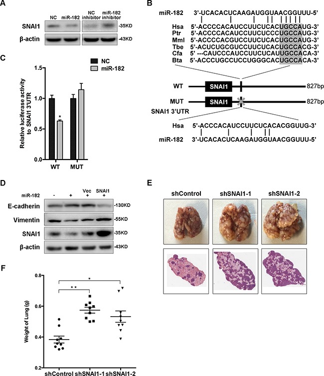
A. Western blot analysis of the protein levels of SNAI1 in MCF-10A cells transfected with miR-182 mimics or inhibitor. β-actin is shown as a loading control. B. A description of predicted miR-182 binding site within the SNAI1 3′UTR, as well as sequence alignment of the miR-182 binding site (WT) and its mutation (MUT) with the miR-182 targeting sequence. C. Luciferase reporter assay of HEK293T cells co-transfected with miR-182 mimics and either WT or Mut luciferase plasmid. D. ‘Rescue’ experiment was performed in MCF-10A cells transfected with NC or miR-182 mimics together with a control (Vec) or SNAI1 plasmid. The protein level of SNAI1 was analyzed using Western blot. β-actin is shown as a loading control. E. SNAI1 expression was blocked using two different targeting sequences in 4T1 cells, then injected into female Balb/c-nu mice via tail vein, respectively. Three weeks later, the lungs were excised and fixed in bouins fixative. Representative gross lungs (upper) and H&E stained lung sections from each group were shown. F. Measurement of weight of excised lungs, representing the macrometastases to lung. Mann-Whitney U-test was used to analyze the significance. Error bars in C and F represent mean ± SEM. *P<0.05, **P<0.01.