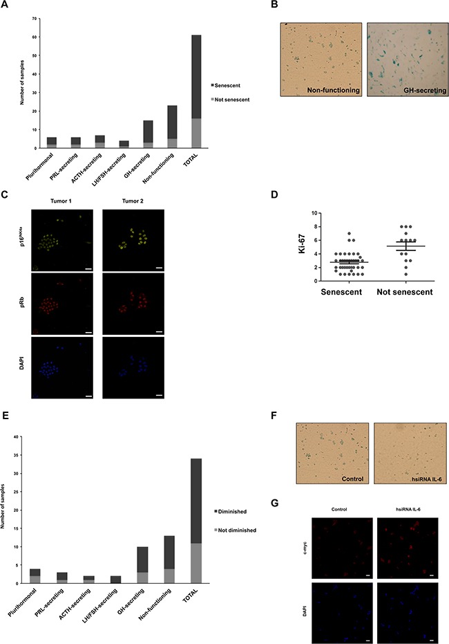
Figure 3. Human pituitary tumor samples express SA-b-gal modulated by IL-6.

A. Primary human pituitary cell cultures prepared from 61 samples of human pituitary tumor samples were stained for SA-β-gal (6 plurihormonal, 6 PRL-secreting, 7 ACTH-secreting, 4 LH/FSH-secreting, 15 GH-secreting and 23 non-functioning tumors). Black zone of the bars represents positive samples, while grey zone represents negative samples. B. Photographs of positive SA-β-gal staining from a non-functioning and a GH-secreting tumor are shown. C. Photographs of positive p16INK4a (yellow; 1:50) and pRb (red, 1:50) staining from a non-functioning and a GH-secreting tumor are shown. 4′,6-diamidino-2-phenylindole (DAPI) were used for staining cell nuclei (blue). Images were acquired at x63 magnification (scale bar: 20μm). D. By Pearson correlation analysis it was determined that senescence correlates negatively with the cellular marker of proliferation Ki-67 in human pituitary adenomas (Pearson coefficient=-0.3245, p<0.05). E. Positive samples of human pituitary tumors (N=34) were electroporeted with 10mM hsiRNA IL-6 or 10mM siRNA GL3 and then stained for SA-β-gal. Black zone of the bars represents samples in which SA-β-gal staining were significantly diminished (Student t test, p<0.05), while grey zone represents samples in which SA-β-gal staining did not vary. F. Photographs of a representative non-functioning tumor with significant decrease in SA-β-gal with hsiRNA IL-6 are shown. Images were acquired at x40 magnification. G. Photographs of a representative non-functioning tumor with significant increase in c-myc (red, 1:50) with hsiRNA IL-6 are shown. 4′,6-diamidino-2-phenylindole (DAPI) were used for staining cell nuclei (blue). Images were acquired at x40 magnification (scale bar: 20μm).
