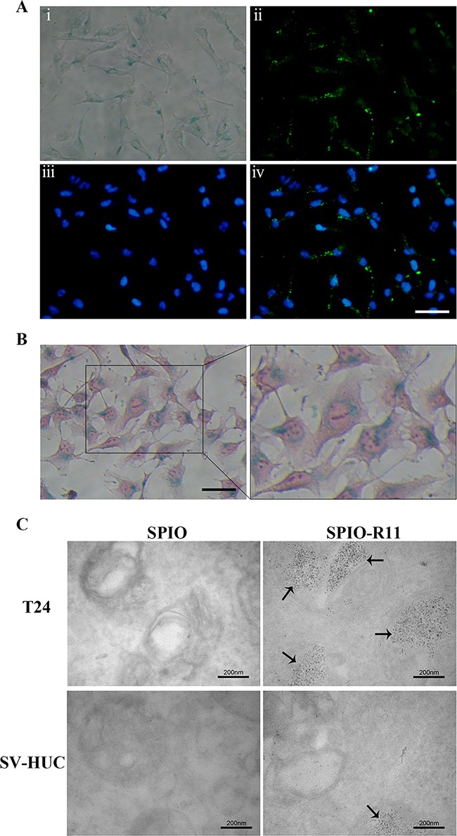
Figure 3. Localization of nanoparticles in T24 cells.

T24 cells were incubated with nanoparticles at an iron concentration of 50 μg/mL for 4 h. A. T24 cells were incubated with SPIO-R11 and then washed with PBS three times. T24 cells were fixed and developed using Prussian blue staining to visualize the presence of iron-oxide nanoparticles; fluorescent images showing FITC, the nuclear counterstain DAPI and merge respectively. FITC was visible by clusters of intense green fluorescence. Scale bar, 50 μm. B. The optical microscope images show 400× imaging of Prussian blue staining and nucleus fast red-counterstained and a magnified view of the black outlines the area. The majority of the blue granules can be seen inside the cytoplasm of the cell. Scale bar, 50 μm. C. TEM images of T24 cells and SV-HUC cells incubated with SPIO and SPIO-R11. A strong uptake of SPIO-R11 was observed and the quantity in T24 cells was much more compared to that in SV-HUC cells, whereas there was no significant uptake for SPIO particles. SPIO-R11 can be seen in vesicles and lysosome of cells. Magnification was at 100,000×.
