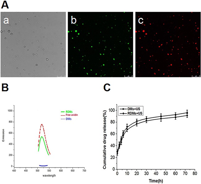
Figure 3. Microbubble fluorescence and drug release characterization.

RDM imaging was performed using CLSM as follows: bright field Aa., dylight488-avidin on RDMs (green fluorescence) Ab., encapsulated DOX in RDMs (red fluorescence) Ac. (scale bar = 25μm). Fluorescence absorbance of RDMs B. DMs and free avidin were examined to assess rituximab conjugation efficiency. In vitro US-triggered DOX release from DMs and RDMs C.
