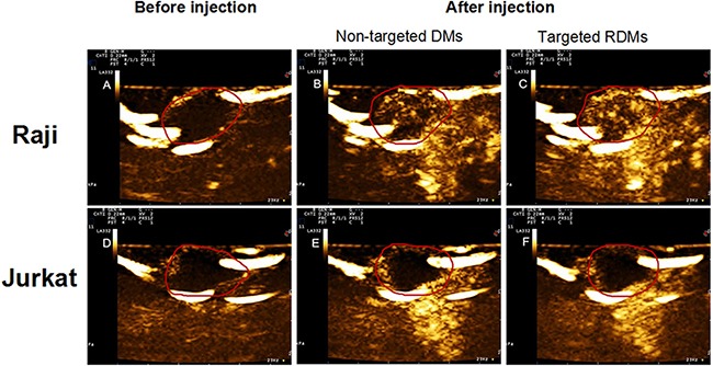
Figure 9. Contrast-enhanced images of targeted RDMs and non-targeted DMs at time to peak in Raji and Jurkat cell-grafted mice.

Images of lymphoma before injection A. & D, non-targeted DMs B. & E. and targeted RDMs C. & F. at time to peak were acquired in Raji and Jurkat cell-grafted mice. RDM peak intensities and contrast enhancement durations were higher than those of non-targeted DMs in Raji cell-grafted mice, and were higher in Raji as compared to Jurkat cell-grafted mice.
