
Figure 1. SkQ1 induced reorganization of cytoplasmic actins and inhibited proliferation of cervical cancer cells.
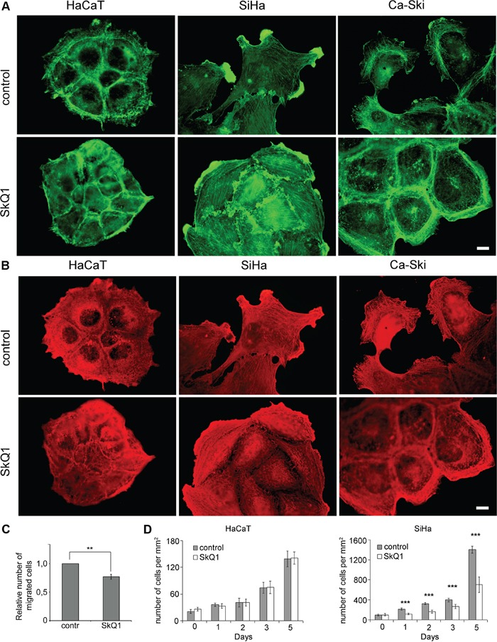
A. β-cytoplasmic actin in the control or cells treated with SkQ1 (40 nM for 3 days). B. γ-cytoplasmic actin in the control or cells treated with SkQ1 (40 nM for 3 days); immunofluorescence microscopy, scale bar 10μm. C. Transwell migration assay performed on SiHa treated with SkQ1 (40 nM), time point 16 hours. Statistically significant difference of data by Mann–Whitney test is marked (**) for p < 0.01. D. Influence of SkQ1 (40 nM) treatment on SiHa and HaCaT cells proliferation; phase contrast microscopy. Statistically significant difference of data by Student t-test is marked (***) for p < 0.001.