
Figure 2. Analysis of EMT markers: cytofluorimetric analysis of Du145 A. and PC3 B. Gene expression of EMT markers after 2 weeks of treatment with or without TGF-ß in DU145 C. and PC3 D.
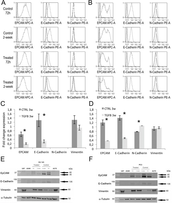
Western blot analysis of EMT markers in Du145 E. and PC3 F. cells after TGF-β treatment for 72h and 2 weeks. Protein lysates of U87 (human glioblastoma cell line), A549 (human lung adenocarcinoma cell line) and A375 (human melanoma cell line) were used as positive or negative controls.