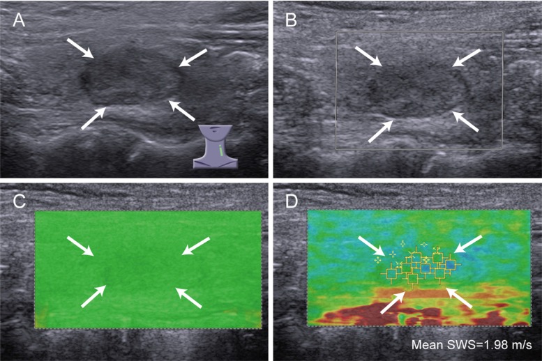
Figure 2. Images of high QM in benign thyroid nodule.
A 54-year-old woman has nodular goiter. (A) conventional ultrasound shows a 14 mm thyroid nodule (arrows) in left thyroid lobe, which is solid, hypoechoic and regular; (B) color Doppler ultrasound shows no color blood flow signal in the nodule (arrows); (C) SW-quality map shows almost green in the nodule (arrows), indicating high QM; (D) the mean SWS of the nodule (arrows) is 1.98 m/s on SW-velocity map.

