
Figure 13. NPM1 is partially involved in TGF-β-mediated EMT.
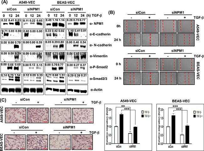
A. A549-Vec and BEAS-Vec cells were treated with NPM1 siRNA (500 nM) prior to administration with TGF-β1 (5 ng/mL) for 24 h. Intracellular expression of NPM1, E-cadherin, N-cadherin, vimentin, phospho-Smad2 and Smad2/3 was detected by immunoblotting. Image intensity was analyzed using ImageJ program (http://rsbweb.nih.gov./ij/plugins). B. Cell migration was performed by a wound healing assay using A549-Vec and BEAS-Vec cells co-treated with NPM1 siRNA and TGF-β1. The assay was repeated twice. C. An invasion assay was performed with A549-Vec and BEAS-Vec cells co-treated with NPM1 siRNA and TGF-β1. Scale bar indicates 100 μm. The assay was repeated twice. Each assay was performed in triplicate, and error bars indicate SD. (**; p< 0.01, ***; p< 0.001).