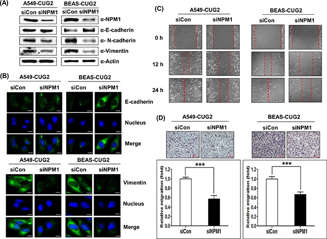
Figure 2. NPM1 silence inhibits the CUG2-induced EMT.

A. At 48 h post-treatment with NPM1 siRNA (500 nM), expression of NPM1, E-cadherin, N-cadherin, and vimentin in A549-CUG2 and BEAS-CUG2 cells was detected by immunoblotting. (siCon; control siRNA, siNPM1; NPM1 siRNA) B. A549-CUG2 and BEAS-CUG2 cells were incubated on chamber slide followed by fixation and permeabilization at 48 h post-treatment with NPM1 siRNA(500 nM), Expression of E-cadherin and vimentin was detected by immunofluorescence using Alexa Fluor 488-conjugated goat anti-mouse IgG (green) and Alexa Fluor 488-conjugated donkey anti-goat IgG (green), respectively. For nuclear staining, DAPI was added prior to mounting in glycerol. Scale bar indicates 10 μm. C. Cell migration was measured by a wound healing assay in A549- CUG2 and BEAS-CUG2 cells at 48 h post-treatment with NPM1 siRNA. The wound closure areas were monitored by phase-contrast microscopy at a magnification of 100×. The assays were repeated twice. D. An invasion assay was performed with A549-CUG2 and BEAS-CUG2 at 48 h post-treatment with NPM1 siRNA. Scale bar indicates 100 μm. The assays were repeated twice. Each assay was performed in triplicate and error bars indicate SD (***; p< 0.001).
