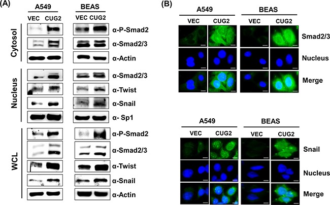
Figure 3. Overexpression of CUG2 activates TGF-β signaling.

A. Expression of phospho-Smad2, Smad2/3, Snail and Twist in A549-CUG2 and BEAS-CUG2 cells was compared with those in their control cells by immunoblotting. In addition, the cells were fractionated into cytosolic and nuclear extracts. Expression of the same proteins was detected by immunoblotting. Sp1 and actin were used loading controls for nuclear and cytosolic extracts, respectively. B. A549-Vec, A549-CUG2, BEAS-Vec and BEAS-CUG2 cells were incubated on chamber slide followed by fixation and permeabilization. Expression of Smad2/3 or Snail was detected by immunofluorescence using Alexa Fluor 488-conjugated goat anti-rabbit IgG (green). For nuclear staining, DAPI was added prior to mounting in glycerol. Scale bar indicates 10 μm.
