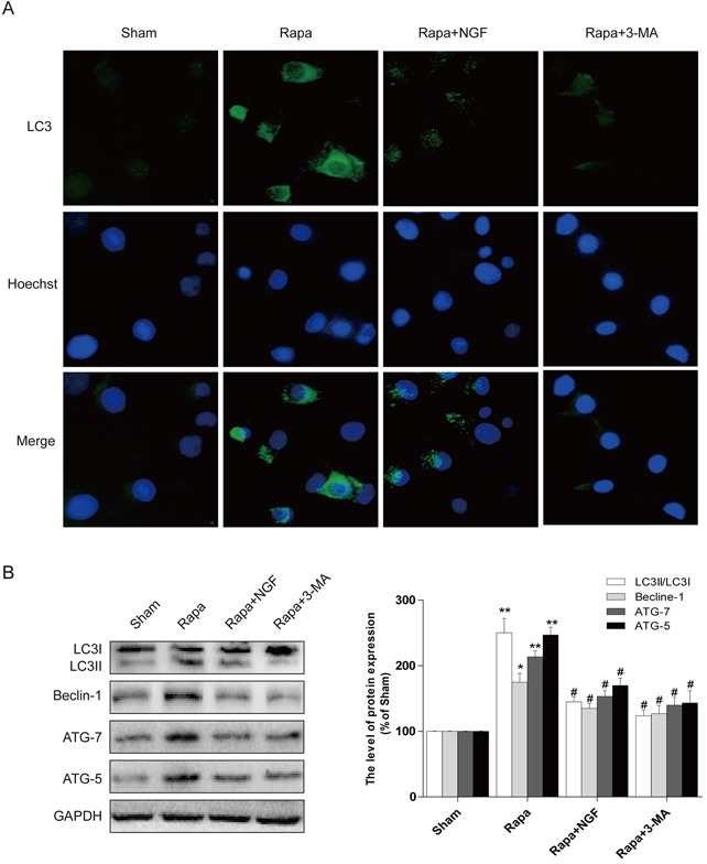
Figure 6. NGF protected H9C2 cells from rapamycin-induced apoptosis through inhibiting autophagy.

A. Representative immunofluorescent staining results of LC3 (marked as green) in H9C2 cells, the nuclei marked as blue with hoechst. B, C. the autophagy related proteins expression in H9C2 cells. The optical density image analysis of autophagy related proteins. * P < 0.05 versus the sham group, # P < 0.01 versus the rapamycin group.
