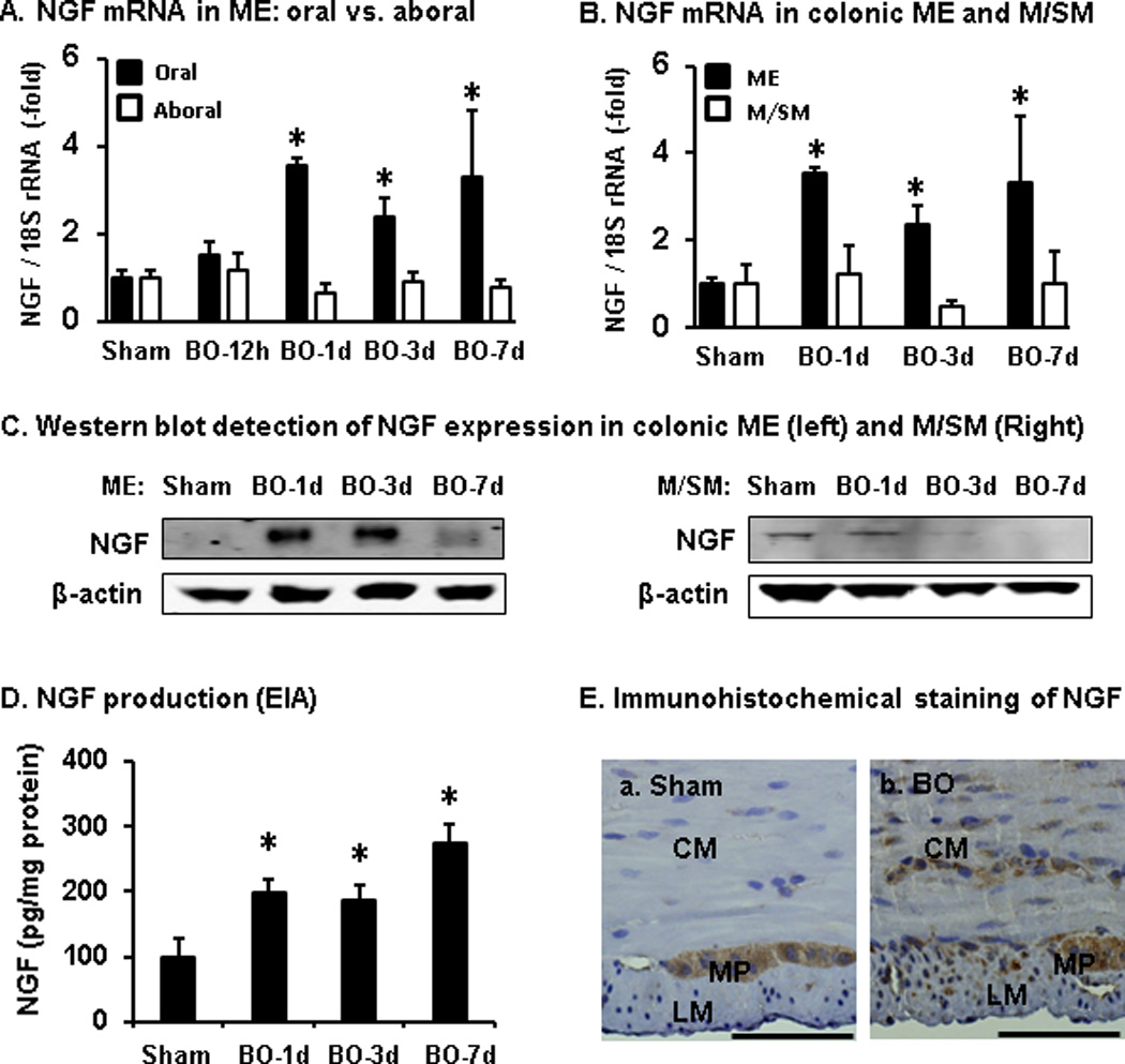
Fig. 3.

Expression of NGF mRNA and protein in the colon in bowel obstruction (BO) detected by qPCR (A, B), Western blot (C), enzyme immunoassay (EIA, D), and immunohistochemistry (E). Note that NGF expression was up-regulated in the distended oral, but not non-distended aboral segment (A, n = 5 each group). The increased NGF expression in the distended colon occurred in the muscularis externae (ME), but not mucosa/submucosa layer (M/SM) (B–D, n = 5 or 6 in each group). Immunohistochemical staining of NGF (in brown, E) in the ME of sham colon (a) and in BO (day 7, b). Bar, 50 µm). CM, circular muscle; LM, longitudinal muscle; MP, myenteric plexus. Images are representative of 4 sham and 4 BO samples. *P < 0.05 vs sham.
