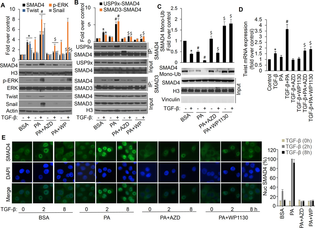
Figure 2.
ERK activation is responsible for FFA promotion of TGF-β-induced USP9x-SMAD4 interaction, SMAD3-SMAD4 complex formation, nuclear SAMD4 retention, and gene expression. MCF-7 cells were treated with BSA or PA in the presence or absence of AZD6244 or WP1130 for 4 h followed by treatment with or without 3 ng/ml of TGF-β for another 4 h. A, Nuclear extracts were made to analyze nuclear SMAD4 levels. Whole-cell extracts were prepared and subjected to Western analysis using p-ERK, ERK, twist, snail and actin antibodies. B, nuclear extracts were made and coimmunoprecipitation of endogenous SMAD4 with USP9x or SMAD3 were performed. Data are presented as mean fold increases (±SD) in treated groups over basal values from three independent experiments. *p<0.01 vs controls; #p<0.01 vs BSA/+TGF-β; $p<0.01 vs PA/+TGF-β. C, nuclear extracts were made and SMAD4 monoubiquitination was detected as described above. *p<0.01 vs controls (BSA/−TGF-β); #p<0.01 PA vs BSA; $p<0.01 PA+AZD vs PA, PA+WP1130 vs PA. D, total RNA was extracted and analyzed for twist mRNA by real-time PCR. *p<0.05 vs controls; #p<0.01 vs TGF-β; $p<0.01 vs PA/+TGF-β. E, MCF-7 cells were treated with TGF-β in the presence or absence of PA, AZD6244 or WP1130 and processed for immunofluorescence with anti-SMAD4 antibody. The same cells were also stained with DAPI to visualize nuclei. Intensity of nuclear SMAD4 among these cells was quantified with Image-Pro Plus 6.0 software. The percentages of nuclear SMAD4 levels illustrated at the right panel represent the mean of three independent experiments, and error bars indicate the SD.

