Figure 3.
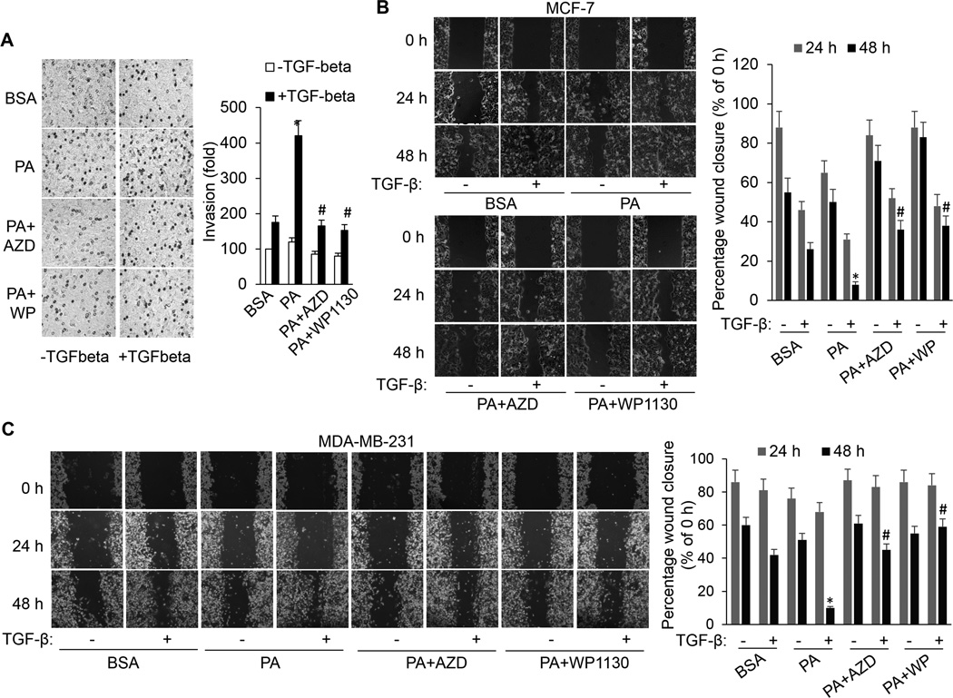
FFA promotes TGF-β-induced invasion and migration by activating ERK and USP9x. A, In vitro invasion assay performed on MCF-7 cells that were treated with BSA or PA in the presence or absence of AZD6244 or WP1130 with or without TGF-β1 for 20 hours. Each column (right panel) represents the mean (± SD) results of two independent experiments. *p<0.05 versus BSA; #p<0.05 versus PA. (B) MCF-7 and (C) MDA-MB-231 cells were scratched with a 20-µl pipette tip and then incubated with BSA, PA, PA plus AZD6244 or WP1130 in the absence or presence of TGF-β1. Migrating cells were photographed under a phase contrast microscope. The percentage of the wound closed was quantified from three independent replicates and is expressed as mean ± SD. *p<0.05 vs BSA/TGF-β; #p<0.05 vs PA/TGF-β.

