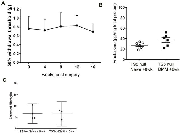
Fig 3.
A) Mechanical allodynia was assessed in the ipsilateral hind paw of Adamts5 null mice (n=5) through 16 weeks after DMM surgery. No statistical differences from time 0 were detected; B) DRG cells were cultured from Adamts5 null mice, naïve or 8 weeks after DMM, and supernatants were analyzed for fractalkine protein. Dots represent individual culture wells. p=0.09; C) Numbers of activated microglia were counted in the ipsilateral and contralateral L4 dorsal horn 16 weeks after DMM and in naïve control Adamts5 null mice. Each dot represents the mean count of 3 sections for one mouse. mean ± 95%CI.

