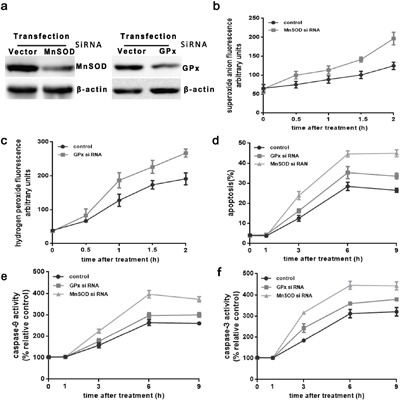
Figure 4. Superoxide mediates induction of the mitochondrial apoptotic pathway in MnSOD and GPx siRNA transfectant HUVEC cells.

HUVEC cells were stably transfected with scrambledsiRNA(Scr) or MnSOD si RNA and GPx si RNA. a. Western blot analysis of MnSOD and GPx protein expression (cropped) in transfected cells. β-actin served as an internal control. b. and c. transfected cells were cultured at 43°C for 2h, and incubated at 37°C for different times as indicated (0h, 0.5h, 1h, or 2h). Superoxide anion assay kit and PF6-AM analysis heat stress-induced O2.- and H2O2, respectively. d-f. Transfected cells were cultured at 43°C for 2h, then further incubated at 37°C for different times as indicated (0h, 1h, 3h, 6h, or 9h). Apoptosis was analyzed by flow cytometry using Annexin V-FITC/PI staining. Enzymatic activity of caspase-9 and-3 was measured in cell lysates using the fluorogenic substrates Ac-LEHD-AFC and Ac-DEVD-AMC, respectively, and activity was expressed relative to the control at 37°C. Each value represents the mean ± SD of three separate experiments.
