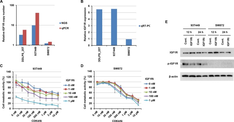
Figure 4. Effects on 93T449 and SW872 cells of combined treatment with CDK4 and IGF1R inhibitors.
(A and B) IGF1R amplification and expression in 93T449 and SW872 cells, as well as an IGF1R-amplified tumor (DDLPS_25T). Relative copy number was estimated by NGS and quantitative PCR (qPCR) (A). mRNA expression was estimated by quantitative RT-PCR (qRT-PCR) and normalized to GAPDH expression (B). Relative expression levels are expressed as ratios of the median expression in non-amplified tumor samples, as in Figure 2B. (C and D) Growth inhibitory effects of CDK4 and IGF1R inhibitors on 93T449 (C) and SW872 (D) cells. Palbociclib (CDK4 inhibitor) and NVP-AEW541 (IGF1R inhibitor) were added at various concentrations, and cell metabolic activities were assayed after 6 days of culture. In this assay, synergism of these inhibitors was evaluated using CompuSyn (http://www.combosyn.com) [41]. Their effects were synergistic in 93T449 cells (average combination index score = 0.42 ± 0.19), but not in SW872 cells (average combination index score = 6.28 ± 3.22). (E) Effect of IGF1R inhibitor on IGF1R phosphorylation in 93T449 and SW872 cells. Cells were treated with NVP-AEW541 (1 μM) for 12 or 24 h and harvested. Expression and Y1135 phosphorylation of IGF1R were evaluated by western blotting analysis.

