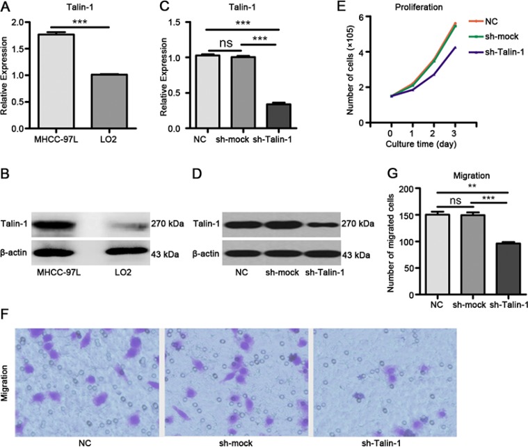
Figure 1. Talin-1 promotes HCC growth and metastasis.
Talin-1 is more highly expressed in MHCC-97L cells compared with LO2 cells in terms of (A) mRNA levels and (B) protein amounts. Talin-1 expression is lower in the sh-Talin-1 group than the NC and sh-mock groups in (C) mRNA levels and (D) protein amounts. (E) Cell proliferation was assessed by counting. Markedly fewer cells were found in the sh-Talin-1 group compared with NC and sh-mock groups at 24 h, 48 h or 72 h; (F, G), HCC cell migration ability was assessed by transwell migration assay. Migration was significantly reduced in the sh-Talin-1 group compared with the NC and sh-mock groups. *P < 0.05 **P < 0.01, and ***P < 0.001. The data are presented as the mean SEM.

