
Figure 5. Effect of 5-FU on hepatic steatosis and inflammation in mice.
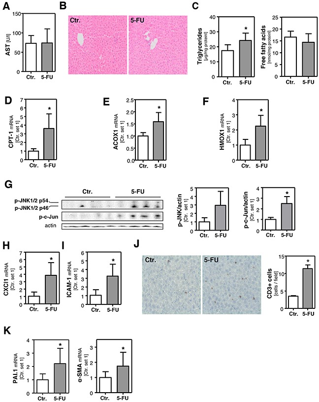
Mice were intraperitoneally injected with a single dose 5-FU (200 mg/kg) and liver tissue samples were collected 24 h after injection. Control mice were injected with solvent isotonic saline solution. A. Plasma levels of aspartate transaminase (AST). B. H/E staining of liver tissue samples. C. Hepatic triglycerides and free fatty acids content normalized to total protein. Analysis of mRNA levels of D. CPT-1, E. ACOX1 and F. HMOX1 by quantitative RT-PCR. G. Western blot analysis of hepatic p-JNK and p-c-Jun protein levels (left panel). Densitometric analysis of p-JNK/actin ratio (middle panel) and p-c-Jun/actin ratio (right panel). Analysis of mRNA levels of H. CXCl1 and I. ICAM-1 by quantitative RT-PCR. J. CD3 staining of liver tissue samples (left panel). Quantification of CD3-positive cells (right panel). Analysis of mRNA levels of K. PAI-1 and α-SMA by quantitative RT-PCR (*: p<0.05 compared to control).