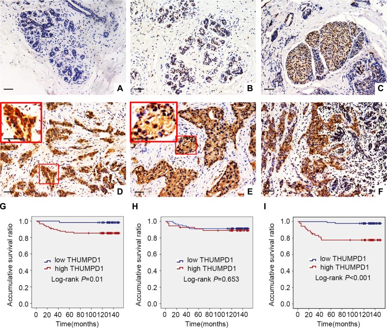
Figure 1. THUMPD1 expression and subcellular localization in breast tumors, and association with patient survival.
THUMPD1 subcellular localization as shown by immunohistochemistry. In normal breast ductal cells, THUMPD1 expression was either absent (A) or weak (B). In carcinoma in situ (C) and IDC cells (D), THUMPD1 was observed in the nucleus and cytoplasm at moderate and high levels, respectively. In some IDC specimens, THUMPD1 was exclusively localized in the cytoplasm (E). THUMPD1 cytosolic and nuclear expression was higher in IDC (F) than in normal breast ductal cells. Magnification, ×200 and ×400. Kaplan-Meier analysis demonstrated that patient overall survival negatively correlated with overall (G) and cytosolic (H), but not nuclear (I), THUMPD1 expression.

