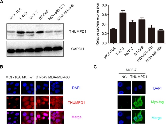
Figure 2. THUMPD1 expression and subcellular localization in breast cancer cell lines.
THUMPD1 expression was higher in most tested breast cancer cell lines compared to normal breast cells (MCF-10A), but was lower in MDA-MB-231 and MDA-MB-468 cells (A). Immunofluorescence analyses indicated that THUMPD1 was mainly localized in MCF-10A cell nuclei, and in both nuclei and cytoplasm of breast cancer cells (B). Immunofluorescence analysis of THUMPD1-myc expression in MCF-7 cells using a Myc-tag antibody (C). Overexpressed THUMPD1 was predominantly localized in the cytoplasm. No positive signal was detected in untransfected cells. Magnification, ×600.

