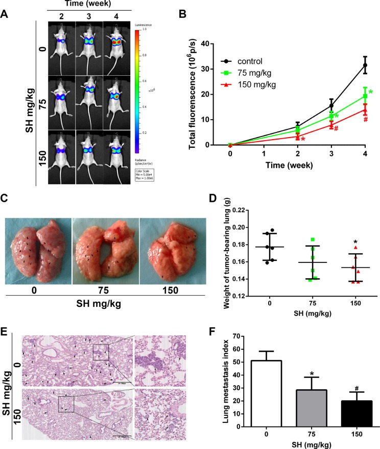
Figure 2. SH reduced pulmonary metastasis in an MDA-MB-231-luc experimental metastatic mouse model.
BALB/c mice were injected with MDA-MB-231-luc via tail vein. (A) Representative bioluminescence images of lung metastasis. Luciferin was injected i.p. and lung metastasis was monitored by a Xenogen IVIS 2000 Imager at the indicated time points. The color scale to the right of the image showed the intensity range of photon flux per second. (B) The quantification of total lung fluorescence. (C) Representative pictures of gross lung. Black arrows indicated metastatic nodules. (D) Lung weight. (E) HE staining of lung specimen and (F) lung metastasis index. Data are represented as mean ± S.D. of three independent experiments. *P < 0.05, #P < 0.01, SH treated group compared with the untreated control group.

