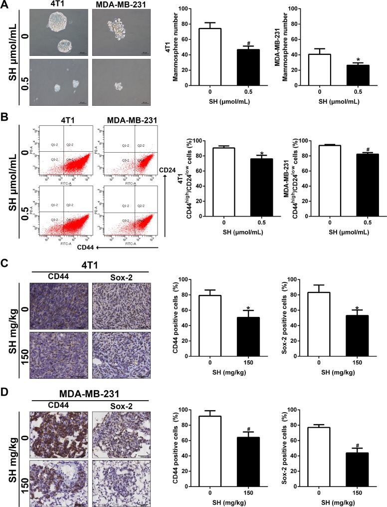
Figure 6. SH inhibited CSC characteristics in breast cancer cells.
(A) Mammosphere formation and its quantification. 4T1 and MDA-MB-231 were treated with SH for 48 h, harvested and seeded on ultra-low attachment culture plates. Mammospheres with diameter > 50 μm were counted on day 10. (B) CSC markers CD44 and Sox-2 were analyzed by western blot. (C and D) IHC analysis of CD44 and Sox-2 in tumor specimens of 4T1 and lung specimens of MDA-MB-231. Data are represented as mean ± S.D. of three independent experiments. *P < 0.05, #P < 0.01, SH treated group compared with the untreated control group.

