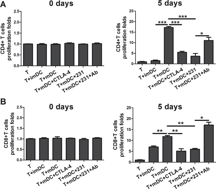
Figure 4. CTLA-4+BCCs inhibit the APC function of mDCs.
Different conditioned DCs (imDC, mDC, mDC+CTLA-4, mDC+231, mDC+231+Ab) were cocultured with allogeneic CD4+T cells (A) or CD8+ T cells (B) at ratio of 1:5, the pure T cell proliferation was also monitored as control. The proliferation was measured by CCK8 method on the day 0 and 5, respectively. The bar graphs represent the ratio of CD4+ T or CD8+ T cells proliferation as mean ± SD of triplicate samples. Results are representative of three independent experiments. *P < 0.05, **P < 0.01, and ***P < 0.001.

