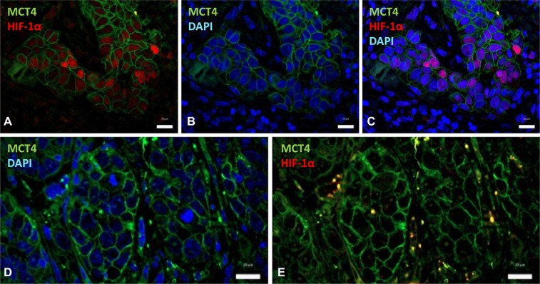
Figure 4. HIF-1α partially overlaps with MCT4 over-expression.
Double immunofluorescence labeling of SCC tissues for HIF-1α (red) and MCT4 (green); cell nuclei were stained with 4′,6-diamidino-2-phenylindole (DAPI) (blue). (A–C) Images show that nuclear-HIF-1α and membrane-MCT4 may co-localize in the same cancer cells. (D, E) Representative images of a different tumor region that contains negative nuclear-HIF-1α and positive membrane-MCT4 immunostainings. Scale bars, 20 μm.

