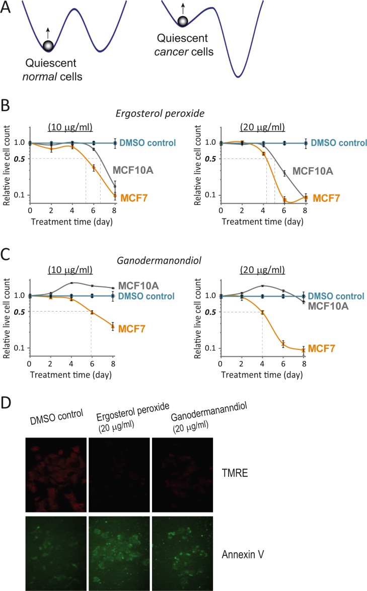
Figure 4. Quiescent MCF7 cells are more sensitive to compound cytotoxicity than quiescent MCF10A cells.
(A) Quiescent states in cancer versus normal cells. Cancer cells featuring autonomous cell proliferation presumably have a shallower and unstable quiescent state compared to normal cells. (B, C) Compound cytotoxicity time course in quiescent MCF7 vs. MCF10A cells. Serum starvation-induced quiescent MCF7 and MCF10A cells were treated with ergosterol peroxide (B) or ganodermanondiol (C) at 10 and 20 μg/ml as indicated. The relative live cell count (y-axis) was determined as in Figure 1B, at the indicated day (x-axis) during the course of treatment. Error bar indicates the standard error of the median (of four replicates). (D) Induced apoptosis in quiescent MCF7 cells. Serum starvation-induced quiescent MCF7 cells were treated with ergosterol peroxide or ganodermanondiol at 20 μg/ml or DMSO vehicle control. Cells were assayed for mitochondrial membrane potential (TMRE) and externalization of inner membrane phospholipids (Annexin V) after 2 days of treatment.

