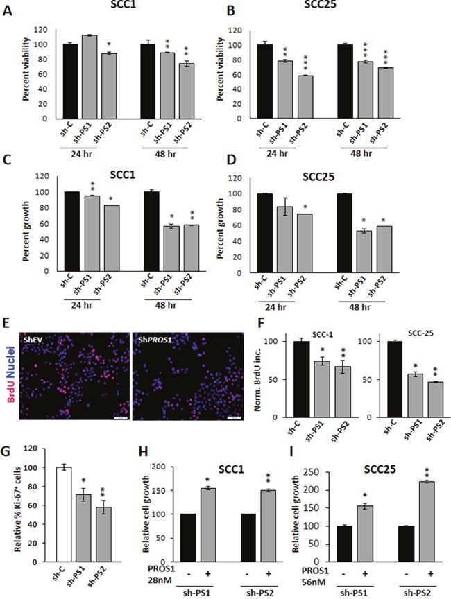
Figure 2. PROS1 kd affects cell viability and proliferation.

A, B. PROS1 knockdown affects cell viability. Analysis of cell viability following PROS1-kd by two different targeting sequences in SCC-1 (A) and SCC-25 (B) cells by the XTT assay. 3,000 cells were seeded in 96 wells, and assayed at the indicated times. Each time point represents the mean ±SEM of at least three replicates. Results are representative of five independent experiments. *P<0.05, **P<0.01; ***P< 0.001. C, D. PROS1 knockdown inhibits OSCC proliferation. Proliferation of control-treated and PROS1-kd SCC-1 (C) and SCC-25 (D) cells as measured at 24 and 48 hours post seeding. Proliferation was measured by crystal violet absorbance, and is presented as relative growth compared to growth of EV-treated cells. Each time point represents 6 replicates in three independent experiments. *P<0.05, **P<0.01. E. BrdU immunohistochemistry (red) indicating decreased BrdU incorporation in PROS1-kd SCC-1 cells compared to shEV. Nuclei are stained with Hoechst (blue). Results are representative of five independent experiments. F. Quantification of BrdU incorporation following PROS1 knockdown in SCC-1 (left) and SCC-25 (right) cell lines. Results are presented as % of BrdU+ cells. Each experiment was performed in N=4 replicates. Five different fields were documented and scored per each condition in every experiment. Results are representative of five independent experiments. *P<0.05, **P<0.01. G. Quantification of Ki-67+ cells. Percent Ki-67+ cells in SCC-25 cells (# Ki-67+/ # Hoechst+ nuclei), normalized to control-treated cells. Each experiment was performed in triplicate, and at least five different fields were scored per condition. Mean values ± SEM are shown from at least three different experiments. *P=0.018; **P= 0.0059. H, I. Exogenous PROS1 rescues the growth rates of PROS1-kd SCC-25 and PROS1-kd SCC-1 cells. PROS1 (28 nmol/L for SCC-1 and 56 nmol/L for SCC-25) was added to the growth medium. Proliferation was measured 48 hours later by crystal violet absorbance, and is presented as relative growth compared to PROS1-kd cells without PROS1. Every experiment was performed in triplicates or sextuplicates. Results are representative of four independent experiments. *P<0.05, **P<0.01.
