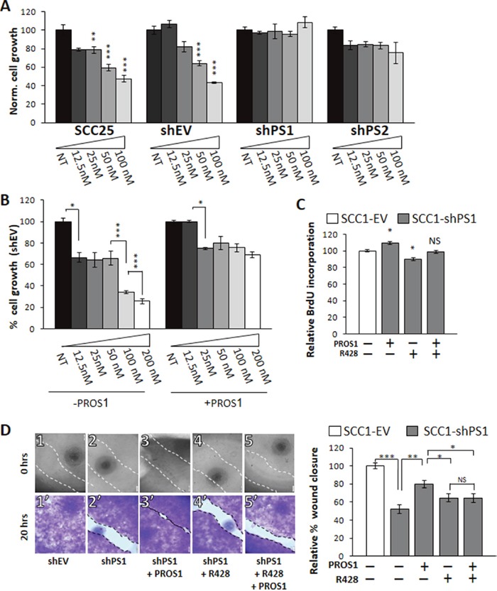
Figure 6. PROS1 modulates the sensitivity of OSCC to the AXL inhibitor R428.

A. PROS1-KD cells are less sensitive to R428. SCC-25 parental, control (shEV), shPS1 and shPS2 cells were subjected to increasing doses of the AXL TKI R248 (12.5-100 nmol/L as indicated) for 48 hours before performing proliferation assays. Proliferation is plotted as a percentage of growth normalized to the number of cells seeded and relative to vehicle-treated cells using the crystal violet method. Parental and control-treated SCC25 cells showed a dose-dependent sensitivity to R428, whereas both sh-PROS1-treated lines lost AXL-mediated R428 sensitivity. The means ± SEM of a representative experiment out of three are shown. *P<0.05, **P<0.01, ***P<0.001. B. Exogenous PROS1 attenuates sensitivity to R428. SCC25-shEV cells were seeded as above, and grown in the presence of R428 dilutions either without or with 28 nmol/L PROS1 for 48 hours. Cell number is plotted as % growth normalized to the number of seeded cells and was detected using the crystal violet method. The means ± SEM of a representative experiment out of three are shown. *P<0.05. C. BrdU incorporation is affected by PROS1 levels. SCC1-shEV and shPS1cells were seeded as described above, and cultured in the presence or not of 28 nmol/L PROS1 and/or R428 (50 nmol/L) for 48 hours. The percent of BrdU+ cells (number of Brdu+ cells/number of nuclei) was calculated. Bars represent the relative mean ± SEM values of one experiment, representative of three independent experiments. *P<0.05, NS = not significant. D. Endogenously expressed PROS1 mediates migration of OSCC through AXL. The % wound area closed 20 hrs after generating the scratch is presented. Inhibition of endogenous PROS1 attenuates wound closure, which is rescued following incubation with PROS1 (28 nmol/L). Inhibition rates obtained for shPS cells that were treated with R428 (25 nmol/L) were similar to those following PROS1 knockdown alone. Scratch images are shown immediately after scratch generation (top panels), and after fixation and staining with crystal violet 20 hours later (lower panels) for each treatment, as indicated. Control-treated cells had closed 100% of the scratch area (1, 1′). By contrast, only 52% of the scratch area was closed by PROS1-sh1 cells (2, 2′). Addition of PROS1 (28 nmol/L) to the growth medium rescued scratch closure (3, 3′). The presence of R428 (25 nmol/L) inhibited scratch closure (4, 4′) and abolished the rescue potential of PROS1 (5′ 5′). Scratch closure measurements are presented as % wound closure. Bars depict the means ± SEM of three independent experiments, with N = 6 replicates in each experiment. ***P< 0.001; **P<0.01; *P<0.05, NS = no statistical significance.
