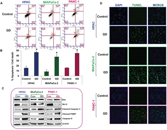
Figure 2. Gedunin induced apoptosis in pancreatic cancer cells.

A. and B. Apoptosis of pancreatic cancer cells treated with 25μM gedunin was detected using flow cytometry. After 24h treatment with Gedunin, cells were stained with Annexin V-FITC/PI. HPAC, MIAPaCa-2, and PANC-1 cells were treated with 25μM gedunin for 24h and protein lysates were subjected to immunoblotting. C. Western blotting was performed using antibodies against Bax, Bcl-2, Cleaved Caspase 3, Cleaved PARP, and Caspase 8. D. Pancreatic cancer cells were treated with 25μM gedunin for 24h and then labeled with DAPI and TUNEL solution. Detection of TUNEL positive cells was observed using confocal microscopy. Data are expressed as the mean ± SEM (*p<0.05) of three separate experiments.
