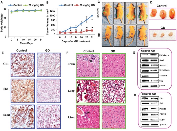
Figure 7. Gedunin inhibited pancreatic cancer growth and metastasis in HPAC xenograft models.

Nude mice bearing HPAC tumors (~100 mm3) were divided into two groups (i) Control (vehicle-DMSO) and (ii) gedunin (20 mg/kg body weight). A. Body weight and B. tumor volume were measured twice weekly for 30 days. C. Control and gedunin treated mice with HPAC xenografts. D. Representative data demonstrating excised tumors of both control and gedunin treated mice. E. Gedunin treated HPAC xenograft tissues were evaluated by IHC for Gli1, Shh, and Snail expression. F. Micrometastasis in control xenografts observed in brain, lung, and liver using hematoxylin and eosin staining. Immunoblots of xenograft tumor tissues for G. EMT markers and H. Hedgehog/Gli1 signaling markers. Data are expressed as the mean ± SEM (*p<0.05) of three separate experiments.
