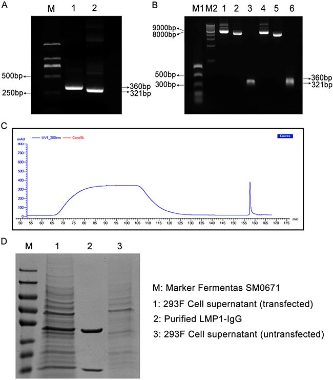
Figure 1.

(A) LMP1-VH and LMP1-VK variable regions were gathered from a previous LMP1-Fab clone. M: Marker DL2000; Lane 1: LMP1-VH variable region (360 bp); Lane 2: LMP1-VK variable region (321 bp). (B) Two recombinant eukaryotic expression vectors (pTH-VH and pTH-VK) were double digested and joined with LMP1-VH and LMP1-VK by Infusion-PCR (IF-PCR). M1: NEB PCR Marker; M2: NEB 1 Kb DNA ladder; Lane 1: pTH-LMP1-VK; Lane 2: Linearized pTH-VK; Lane 3: LMP1-VK; Lane 4: pTH-LMP1-VH; Lane 5: Linearized pTH-VH; Lane 6: LMP1-VH. (C) UV curve of LMP1-IgG purification. (D) SDS-PAGE confirmed the purification of LMP1-IgG. M: Marker Fermentas SM0671; Lane 1: 293F Cell supernatant (transfected); Lane 2: Purified LMP1 IgG; Lane 3: 293F Cell supernatant (untransfected).
