
Figure 3. Immunohistochemistry (IHC) analysis in clinical ENKTL samples.
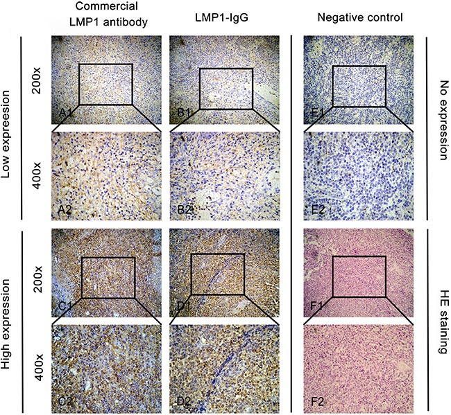
A1 and A2. Low expression of LMP1 when using a commercial LMP1-antibody as the primary antibody in IHC analysis. B1 and B2. Low expression of LMP1 when using LMP1-IgG as the primary antibody in IHC analysis. C1 and C2. High expression of LMP1 when using a commercial LMP1-antibody as the primary antibody in IHC analysis. D1 and D2. High expression of LMP1 when using LMP1-IgG as the primary antibody in IHC analysis. E1 and E2. Negative expression of LMP1 when using phosphate-buffered saline (PBS) in IHC analysis as a negative control. F1 and F2. Hematoxylin-eosin (HE) staining of ENKTL samples. Original magnification: × 200 in A1, B1, C1, D1, E1 and F1; ×400 in A2, B2, C2, D2, E2 and F2.