
Figure 4. LMP1-IgG inhibits proliferation and induces apoptosis of ENKTL cells.
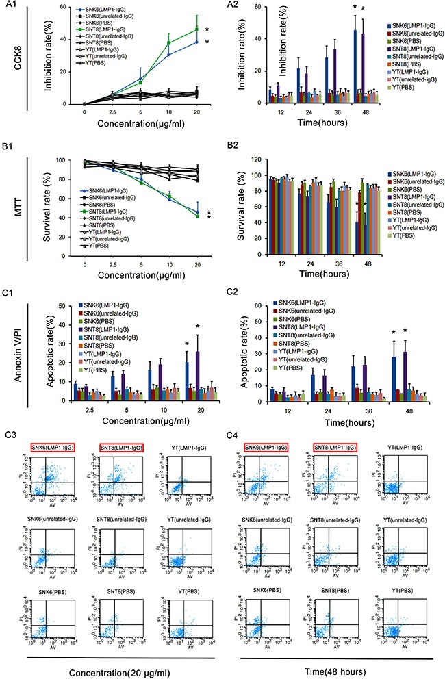
(A1, A2, B1 and B2) CCK8 and MTT assays exhibited the concentration- and time-dependent inhibitory effects of LMP1-IgG (2.5–20 μg/ml or 12–48 h treatment) on the proliferation of SNK6 and SNT8 cells, whereas the inhibitory effect on YT cells was low and insignificant. * Significant difference in SNK6 and SNT8 cells with LMP1-IgG (20 μg/ml or 48 h treatment) compared with PBS treatment. p < 0.05. (C1 and C2) Apoptotic rates in ENKTL cells treated with LMP1-IgG (2.5–20 μg/ml or 12–48 h treatment). *Significant differences in apoptotic rate in SNK6 and SNT8 cells with LMP1-IgG (20 μg/ml or 48 h treatment) compared with PBS treatment. p < 0.05. (C3) Representative images of cell apoptosis, detected with flow cytometry by Annexin V/PI double staining after treatment with LMP1-IgG (20 μg/ml). (C4) Representative images of cell apoptosis, detected with flow cytometry by Annexin V/PI double staining after treatment with LMP1-IgG (48 h treatment). The red frame illustrates the significantly increased apoptotic rate of SNK6 and SNT8 cells treated with LMP1-IgG.