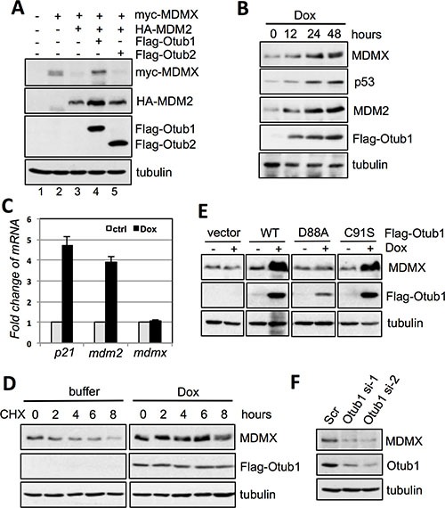
Figure 1. Otub1 stabilizes MDMX and increases its levels.

(A) Otub1, but not Otub2, suppresses MDM2-mediated MDMX degradation. H1299 cells were transfected with the indicated plasmids and assayed by IB. (B) Otub1 induces the levels of endogenous MDMX. T-Rex-U2OS-Flag-Otub1 cells were cultured in the presence of 2 ug/ml doxycycline (Dox) for indicated time points, followed by IB. (C) Otub1 does not induce the levels of MDMX mRNA. T-Rex-U2OS-Flag-Otub1 cells were cultured in the presence or absence of 2 μg/ml Dox for 24 hours, followed by RT-qPCR detection of the relative expression of p21, mdm2 and MDMX mRNA normalized with GAPDH mRNA. (D) Otub1 stabilizes MDMX. T-Rex-U2OS-Flag-Otub1 cells were cultured in the presence or absence of 2 μg/ml Dox for 24 hours. Then the cells were cultured in the presence of cyclohexamide (CHX, 50 μg/ml) for the indicated hours, followed by IB. (E) Otub1 stabilizes MDMX independently of its Dub activity. T-Rex-U2OS-Flag-Otub1 (wt, C91S, or D88A) cells or the control T-Rex-U2OS cells were cultured without or with 2 μg/ml Dox for 24 hours, followed by IB assays. (F) Knockdown of Otub1 reduces the levels of MDMX. U2OS cells were transfected with scrambled or individual siRNA against Otub1 and assayed by IB.
