
Figure 3. Otub1-stabilized MDMX relocalizes to the cytosol and the mitochondria.
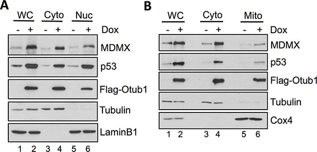
(A) Cell fractionation assays were performed in T-Rex-U2OS-Flag-Otub1 cells treated without or with 2 ug/ml Dox for 24 hour. The cells were fractionated into cytoplasmic (Cyto) and the nuclear (Nuc) fractions, followed by IB with indicated antibodies. (B) Otub1-stabilized MDMX relocalizes to the mitochondria. The cytoplasmic fraction was further separated into the cytosol and mitochondria (Mito) fractions by further centrifugation and assayed by IB using the indicated antibodies. WC: whole cell lysates. Tubulin, lamin B1 and Cox4 were used as the cytoplasm, nucleus and mitochondria markers, respectively.