
Figure 2. miR-138-5p inhibits the autophagy flux in pancreatic cancer cells.
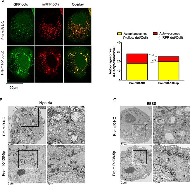
(A) After transfection of PANC-1 cells with Ad-tf-LC3 for 72 h, the cells were transfected with pre-miR-NC or pre-miR-138-5p, and exposed to serum-free medium for 4 h. Representative images of fluorescent LC3 puncta are shown. The mean number of autophagosomes (yellow puncta in merged images) and autolysosomes (red puncta merged images) is shown in the right panel (n = 3). *P < 0.05. (B) Transmission electron microscopy showed autophagosomes (black arrow) in PANC-1 cells of the pre-miR-NC group after hypoxia for 48 h, whereas broken and swollen mitochondria were observed in the pre-miR-138-5p group without autophagic signs. (C) Transmission electron microscopy showed autophagosomes in PANC-1 cells of the pre-miR-NC group after serum-free medium culturing for 12 h. Mitochondria swelling and crista fragmentation were observed in the pre-miR-138-5p group (n = 3).