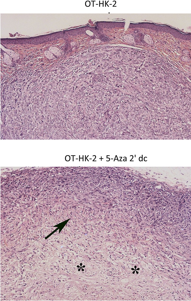
Figure 7. Representative photomicrographs showing histopathological changes in xenograft tumor samples.

Tumor samples from athymic nude mice inoculated with OT-HK-2 cells with and without 5-aza 2’ dc treatment were fixed in 10% neutral buffered formalin, processed for paraffin embedding, sections (5μM) were cut, stained with haemotoxylin and eosin stain and evaluated microscopically for histopathological changes. In 5-aza 2’ dc treated group, decreased tumor volume/growth was histologically characterized by areas of decrease in cellularity (arrows) with prominence of connective tissue portion (asterisk).
