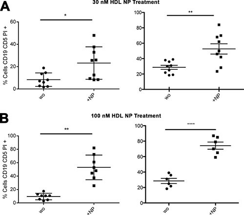
Figure 5. HDL NPs induce apoptosis in isolated human CLL cells.

(A) Percent of apoptotic isolated CLL cells without treatment (wo) compared to cells treated with HDL NPs (30 nM) for 72 hours (USA, left: 8.3% ± 6.1% versus 23.2% ± 14.5%; *p = 0.0059; Italy, right: 28.6% ± 2.6% versus 52.6% ± 6.7%; **p = 0.043). (B) Percent of apoptotic CLL cells without treatment compared to cells treated with HDL NPs (100 nM) for 72 hours (USA, left: 9.5% ± 4.8% versus 53.0% ± 18.4% **p = 0.0008; Italy, right: 28.52% ± 3.3% versus 74.2% ± 4.6%; ****p < 0.0001).
