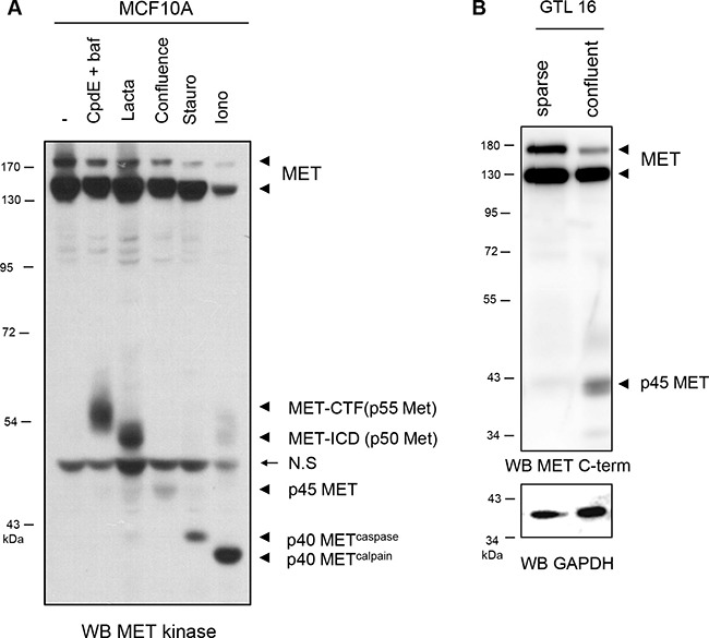
Figure 3. Generation of the MET fragments in epithelial cells.

(A) MCF10A cells were treated for 5 h with 1 μM compound E and 5 nM bafilomycin (CpdE+baf), for 5 h with 10 mM lactacystin (Lacta), for 6 h with 1 μM staurosporine (Stauro), for 1 h with 1 μM ionomycin (Iono), or cultured to high density (confluence). Cell lysates were analyzed by western blotting with an antibody directed against the kinase domain of MET. (B) Sparse and confluent GTL16 cells were cultured 48h. Cell lysates were analyzed by western blotting with an antibody directed against the C-terminal region of human MET (MET C-term) and GAPDH to assess the loading. Arrowheads indicate full-length MET, MET CTF (p55 MET), MET-ICD (p50 MET), p45 MET, the p40 METcaspase and p40 METcalpain. Arrow indicates position of a non specific band (N.S).
