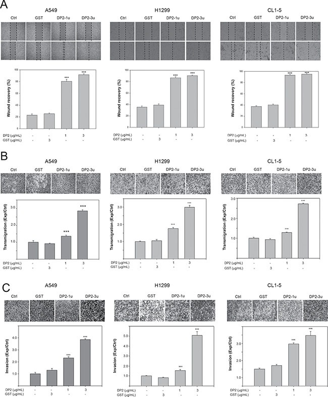
Figure 1. DP2 promotes cell migration and invasion of NSCLC cells.

(A) Wound healing assay was performed in the three cell lines with 48 h of recovery. Cells were cultured on 6-well plates, scratched using tips, and then incubated with GST (3 μg/mL), DP2-1u (1 μg/mL), or DP2-3u (3 μg/mL) for 48 h. After the incubation, wound recovery was determined as comparing to the initial of each treatment group. (B) and (C), cells were subjected to transmigration and invasion assay with incubation of GST (3 μg/mL), DP2-1u (1 μg/mL), or DP2-3u (3 μg/mL) for 24 h. The transmigrated cells on the bottom side of membrane were stained and counted using light microscope at a magnitude of 200X. Transmigration and invasion were presented as the ratio of treatment/control. Ctrl, control; ***P < 0.005 as compared to GST treatment.
