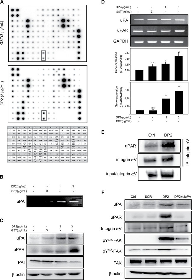
Figure 4. DP2 upregulated uPA/uPAR expression, enhanced uPAR/integrin αV interaction and induced FAK activation.

(A) and (B), cells were incubated with GST or DP2 at 3 μg/mL for 24 h, and then the cultured medium was subjected to cytokine array assay or uPA activity assay. (C) and (D), cells were incubated with GST or DP2 at 3 μg/mL for 1 or 4 h, and then lysed for immunodetection of the indicated proteins or for total RNA extraction and the following mRNA expression analysis using RT-PCR and qRT-PCR. (E) cells were incubated with GST or DP2 at 3 μg/mL for 24 h, and then lysed for immunoprecipitation using antibodies against integrin αV. The precipitated proteins were subjected to immunodetection of uPAR and integrin αV. (F) cells were transfected with scramble RNA (SCR) or specific siRNA against uPA (siuPA), treated with DP2 at 3 μg/mL for 24 h, and then lysed for immunodetection of determining the level of indicated phosphorylated proteins or total proteins. β-actin was sued as internal control.
